Figure 9.
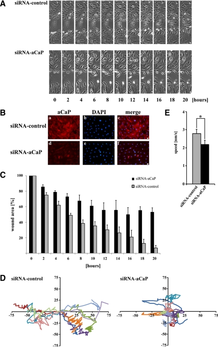
h3/acidic CaP enhances wound healing in REF52.2 cells in an in vitro assay. (A) REF52.2 cells were seeded on glass-bottomed culture plates and either transfected with siRNA against rat h3/acidic CaP (siRNA-aCaP) or with nontargeting control-siRNA the next day. Seventy-two hours after transfection, the cell layer was wounded with a sterile pipette tip, and the healing procedure was observed for 20 h. Bar, 20 μm. (B) To verify h3/acidic CaP knockdown, cells were immunostained after the in vitro wound healing assay for endogenous aCaP expression by using the rabbit polyclonal anti-h3/acidic CaP antibody. Bar, 20 μm. (C) To quantify in vitro wound healing, the wound area was measured using ImageJ software and calculated as a percentage of the wound area at 0 h. Data represent three independent experiments. (D) Ten single cells of each group (left, control cells; right, h3/acidic CaP knockdown cells) were manually traced using ImageJ software and migration tracks are shown as scatter plots. (E) The average speed of 30 individual cells in total, coming from three independent experiments, for either h3/acidic CaP knockdown or control cell groups was calculated and plotted in a graph bar. Note that the difference is statistically significant (p = 0.017).

