Figure 1.
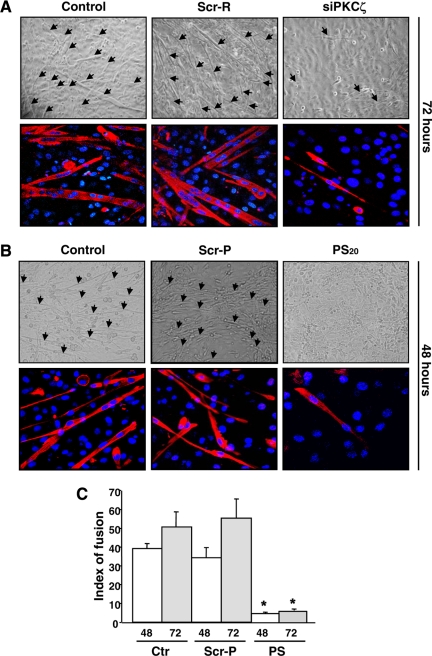
Inhibition of PKCζ expression and activity prevents the formation of myotubes. (A) The down-regulation of PKCζ by siRNA abrogated the myoblast fusion and myotube formation. Primary myoblasts were transfected with PKCζ siRNA (siPKCζ) or scrambled siRNA (Scr-R) and induced to differentiate. At 72 h of differentiation, cells were fixed and double-stained with anti-MHC antibody, and DAPI, to detect differentiated cells and nuclei, respectively. The arrows in the phase-contrast images indicate myotubes. The bottom panels shows fluorescence labeling of MHC (red) and DNA (DAPI, blue), visualized by confocal microscopy. The images reveal a pronounced decrease in cell fusion and myotube formation as well as in MHC expression, when PKCζ is down-regulated. (B) The inhibition of PKCζ activity by a pseudosubstrate inhibitor peptide (PS) blocked the fusion of myoblast to multinucleated myotubes. Mouse primary myoblasts were induced to differentiate in the presence of either the PKCζ inhibitor peptide (PS; 15 or 20 μM) or the scrambled peptide (Scr-P; 20 μM). After 48 h of differentiation, cells were fixed and double-stained with antibodies against MHC and DAPI. Similarly to PKCζ down-regulation by siRNA (Figure 1A), PKCζ inhibition prevented myotube formation. (C) The fusion indexes of mouse primary myoblasts were determined in the presence of both the inhibitor peptide (PS, 20 μM) and the scrambled peptide (Scr-P, 20 μM). The fusion index was determined as a ratio of the number of nuclei in myotubes (showing at least two nuclei) to the total number of nuclei in six randomly chosen microscopic fields. The data are shown as means ± SEM (n = 3; *p < 0.05; Student's t test).

