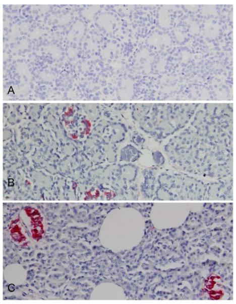
Figure 5. Localization of human insulin within a chimeric sheep pancreas three months after transplantation with human fetal pancreatic mesenchymal stem cells.
Human insulin containing cells were identified by immunohistochemistry using monoclonal anti-human insulin antibody (clone MAB 1). (B) Chimeric sheep no 2361 sacrificed one day after birth display cell clusters with human insulin identified by red coloration.
(A) Age matched non-transplanted negative control animal tissue section stained with the same antibody is negative.
(B) Chimeric sheep no 2361 sacrificed day one after birth displays cell clusters with human insulin secretion identified by red coloration
(C) Human positive control pancreas stained for insulin (red coloration). Original magnifications × 20.

