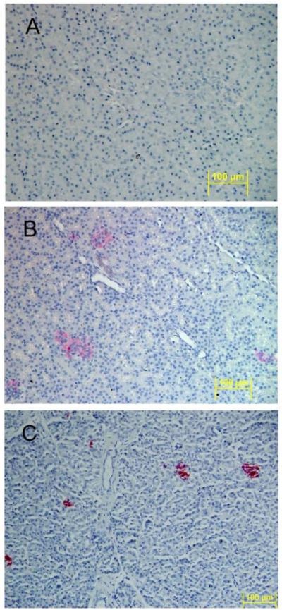
Figure 6. Localization of human insulin within a chimeric sheep pancreas 27 months after transplantation with human fetal pancreatic mesenchymal stem cells.
Human insulin containing cells were identified by immunohistochemistry using human-specific insulin antibody (clone MAB 1).
(A) Age matched non-transplanted negative control sheep pancreas section stained with anti-human insulin is negative for red coloration.
(B) Chimeric sheep sacrificed at 27 months after transplantation stained with anti-human insulin shows cell clusters with red coloration.
(C) Human pancreas stained with anti-human insulin demonstrates cell cluster with red coloration (positive control). Original magnifications × 20.

