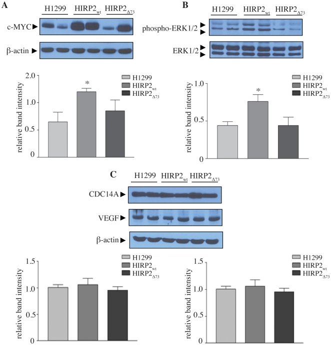
Figure 6. Tumor xenografts derived from HIRP2wt cells display an increase of c-MYC expression and ERK1/2 phosphorylation.
Extracts from tumor tissue were analyzed by Western blotting with antibodies against c-MYC, phospho-ERK1/2, ERK1/2, CDC14A, VEGF and β-actin. Representative immunoblots and quantification from three independent experiments (n = 9 mice)of (A) c-MYC, relative to β-actin; (B) phospho-ERK1/2, relative to ERK 1/2; (C) CDC14A and VEGF, relative to β-actin. Data are expressed as means of relative band intensity ± SEM. * p<0.05 versus H1299 (Student's t-test).

