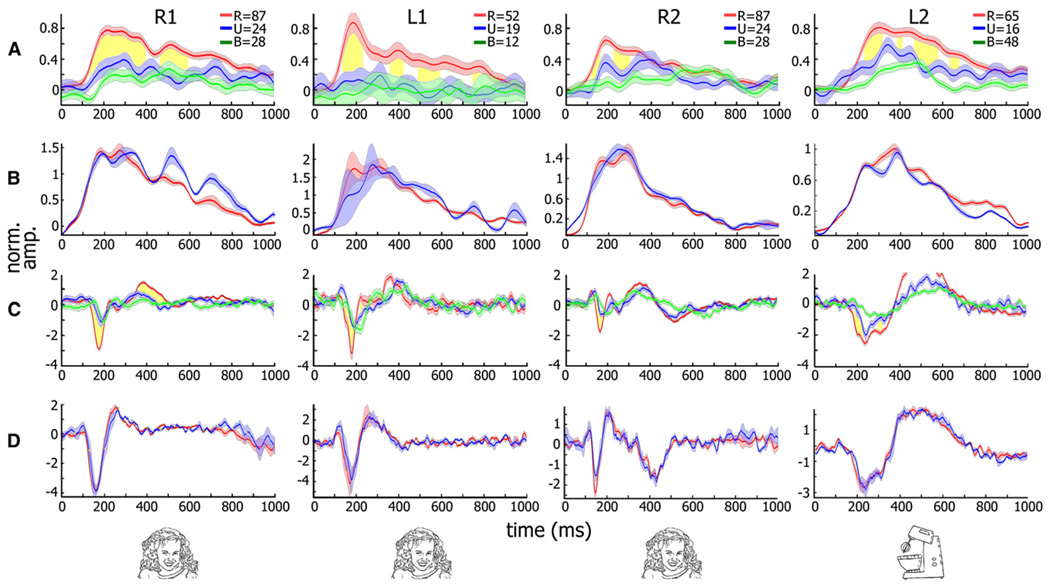
Figure 4. Neuronal Responses of Category-Selective Electrodes in the Backward Masking Experiment.
(A) Gamma BLP (30–70 Hz band) response of four category-selective (three face-selective and one man-made object-selective) electrodes from three patients to 16 ms presentation of targets from the preferred category at the critical SOA (16, 33, 16, and 83 ms from left to right). Average responses when subjects succeeded or failed in recognizing the images are shown in red and blue, respectively. Mask-only responses are shown in green. SEM is given by shading. Time points showing significant recognition-related increase in gamma BLP (p < 0.01, corrected) are shown in yellow.
(B) Control showing responses to the same exemplar composition but at longer SOA in which patients successfully recognized both sets.
(C and D) Same analysis as (A) and (B) but for evoked responses. Numbers of trials are for (A) and (C). R, recognized; U, unrecognized; B, mask-only trials. Note the significant and long-lasting increase in gamma BLP (A) and the increase in the evoked (C) response amplitudes associated with successful recognition for all electrodes shown (see text). No such enhancement was seen when the same image compositions were compared during successful recognition of both sets (B and D).

