
Figure 2. VASP phosphorylation plays a negative role in filopodia/microspikes formation.
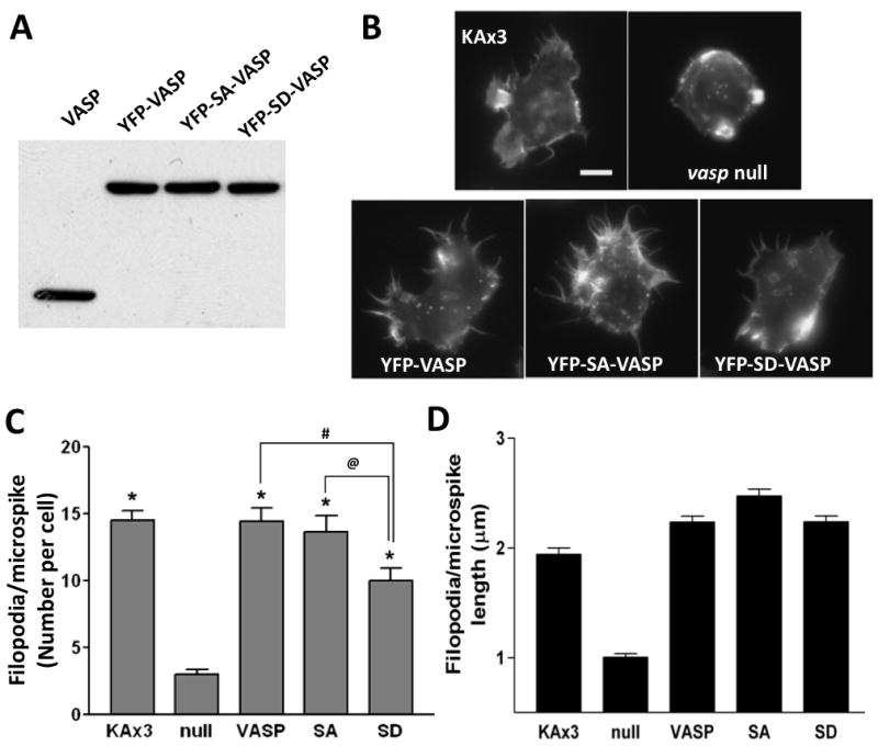
A, Expression of YFP-tagged VASP mutants in vasp null cells. Cells at vegetative stage were lysed and whole cell lysates (30 μg of proteins) were run on SDS-PAGE and subjected to immunoblotting with anti-VASP antibody. Immunoblot shows endogenous (VASP) and exogenous (YFP-VASP) expression. B, vegetative cells were synchronized in Na/K phosphate buffer for 30 min followed by permeabilization, fixation and staining with Texas Red-phalloidin. Scale bar represents 5 μm. C-D, the number (C) and the length (D) of filopodia/microspikes per cell were counted and presented as a bar graph. Data presented as mean ± SEM. Unpaired Student t-test. (* p<0.0001, compared to null cells; # p<0.01; @ p<0.05)