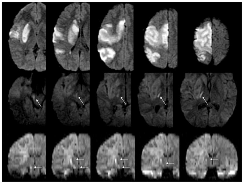
Figure 1.

MRI in the acute and subacute time period. Diffusion-weighted imaging performed immediately after left-sided weakness was recognized shows an acute stroke, infarct volume of 155 cm3 (upper panels); MRI performed 11 days postsymptom recognition shows Wallerian degeneration (arrows) on axial diffusion-weighted images (middle panels) and coronal reconstruction diffusion-weighted images (arrows) in lower panels.
