
Figure 1. Myofibers in a variety of model systems form from the fusion of mononucleated muscle precursors.
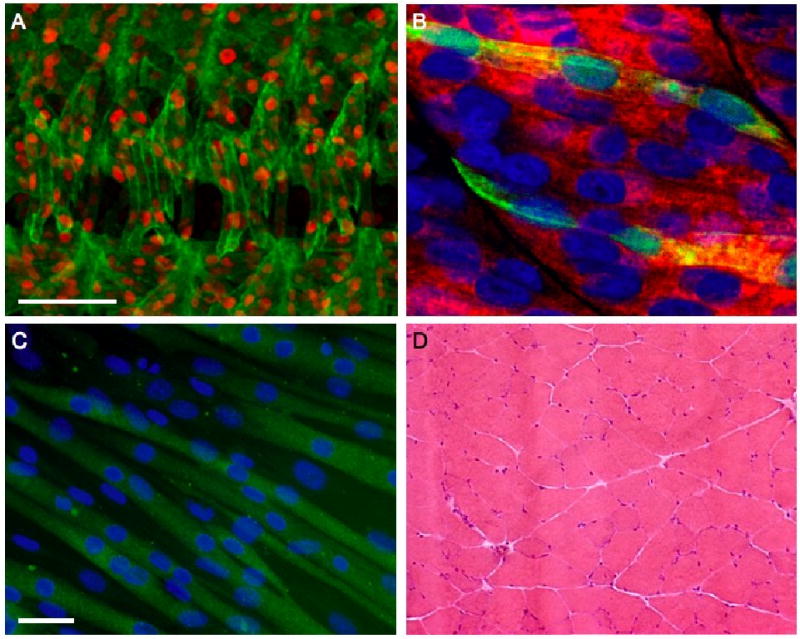
(A-D) Nuclei were visualized using a nuclear DsRed transgene (red) (A), DAPI (blue) (B, C) or hematoxylin (purple) (D). (A) Four hemisegments of a Drosophila embryo were analyzed by immunohistochemistry using antibodies against tropomyosin (green). (B) The syncytial fast-twitch muscle fibers of a wild-type zebrafish embryo were labeled with antibodies against fast myosin light chain (red). Two fast muscle fibers are highlighted by GFP expression (green) from a skeletal muscle actin∷gfp transgene. (C) C2C12 myoblasts, a satellite cell-derived mouse myoblast cultured cell line, were analyzed by immunohistochemistry using antibodies against myosin heavy chain (green). The scale bar represents 40 μm. (D) Cross sections of a major leg muscle, the gastrocnemius, of an adult mouse stained with hematoxylin and eosin. Note that the nuclei are peripherally located and, unlike in other panels, the multinucleate nature of the myofiber is not clearly evident from this section.