
Figure 1.
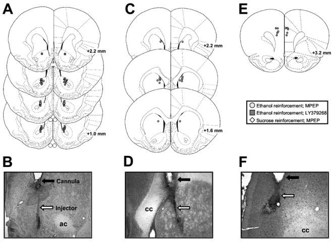
Illustrations of cannulae and injector placements. Injector placements from individual rats with accurate bilateral placements in the (A) nucleus accumbens, (C) dorsomedial caudate, and (E) medial prefrontal cortex. Circles represent animals trained on ethanol self-administration tested with 2-methyl-6-(phenylethynyl)pyridine hydrochloride (MPEP); squares represent animals trained on ethanol self-administration tested with LY379268; and diamonds represent animals trained on sucrose self-administration tested with MPEP. Representative photomicrographs showing cannula (closed arrows) and injector (open arrows) tracks in nucleus accumbens (B), dorsomedial caudate (D), and medial prefrontal cortex (F). ac, anterior commissure; cc, corpus callosum. Figures A, C, and E published in The Rat Brain in Stereotaxic Coordinates, 4th ed. (CD-ROM) by Paxinos and Watson, Copyright Elsevier (1998).