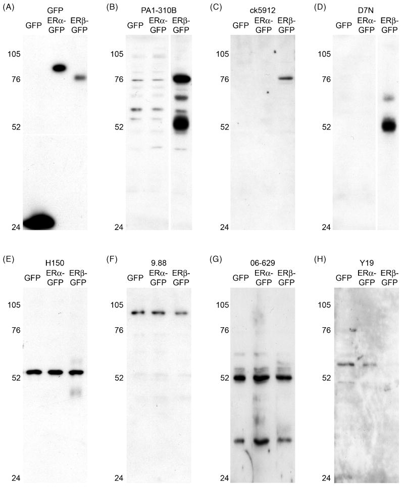
Figure 2.
Detection of ERβ in transfected HT22 cells by western blot. (A-H) Western blots of HT22 cells transfected with GFP, ERα-GFP, or ERβ-GFP and probed with anti-GFP or various anti-ERβ antisera: (A) GFP is detected in all transfected cells, as appropriate. (B) PA1-310B anti-ERβ detected a prominent, appropriately sized band of ∼78 kDa in ERβ-GFP transfected cells, and also a prominent band at ∼52 kDa, which could reflect ERβ cleaved from GFP. PA1-310B also detected faint bands in ERα-GFP transfected cells. (C) ck5912 anti-ERβ detected a single appropriately sized band of ∼78 kDa only in ERβ-GFP transfected cells. (D) D7N anti-ERβ detected a single a band of ∼52 kDa in ERβ-GFP transfected cells, which could reflect ERβ cleaved from GFP. (E) H150 anti-ERβ detected a single band of inappropriate size (∼55 kDa) in all transfected cells. (F) 9.88 anti-ERβ detected a single band of inappropriate size (∼102 kDa) in all transfected cells. (G) Similar to H150, 06-629 anti-ERβ detected a band close to 52 kDa in all transfected cells, as well as band of ∼38 kDa in all transfected cells. (H) Y19 anti-ERβ detected faint bands in GFP- and ERα-GFP transfected cells.

