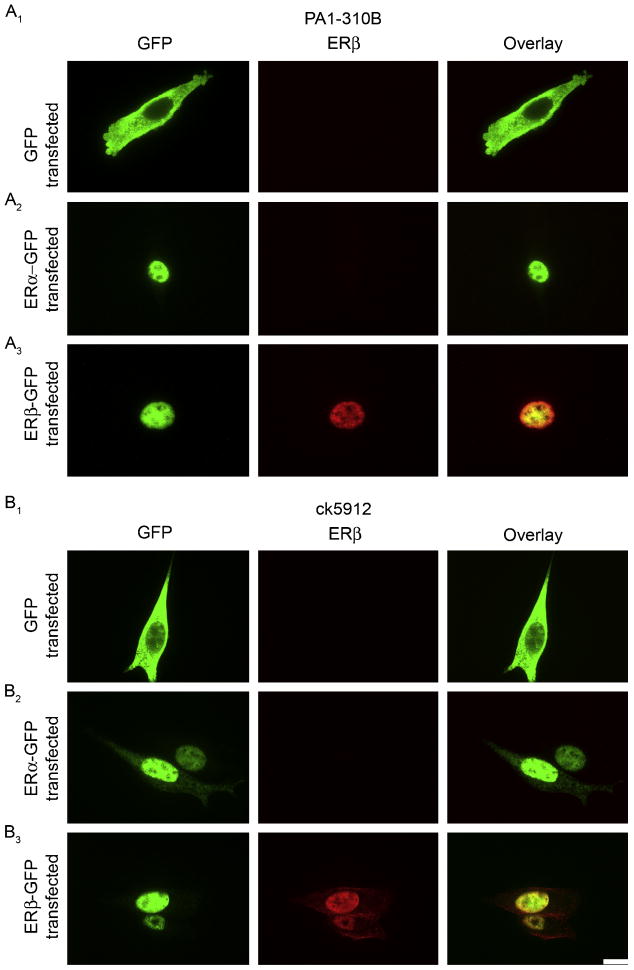
Figure 3.
Detection of ERβ in transfected HT22 cells by immunocytochemistry. (A) Representative images of PA1-310B immunostaining in HT22 cells transfected with (A1) GFP, (A2) ERα-GFP, or (A3) ERβ-GFP. PA1-310B anti-ERβ detected nuclear labeling only in ERβ-GFP transfected cells, as appropriate. (B) Representative images of ck5912 immunostaining in HT22 cells transfected with (B1) GFP, (B2) ERα-GFP, or (B3) ERβ-GFP. Ck5912 anti-ERβ detected nuclear labeling only in ERβ-GFP transfected cells, as appropriate. Scale bar is 10 μm and applies to all frames.

