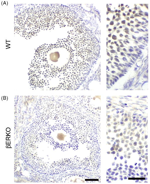
Figure 4.
ERβ immunoreactivity in wildtype (WT) and ERβ knockout mouse (βERKO) ovaries. Representative photomicrographs from (A) WT and (B) βERKO ovaries labeled with PA1-310B anti-ERβ. ERβ immunoreactivity is detected in granulosa cells of both WT and βERKO mice, although less intense in βERKO. Scale bars are 50 μm and 25 μm, left and right panels, respectively.

