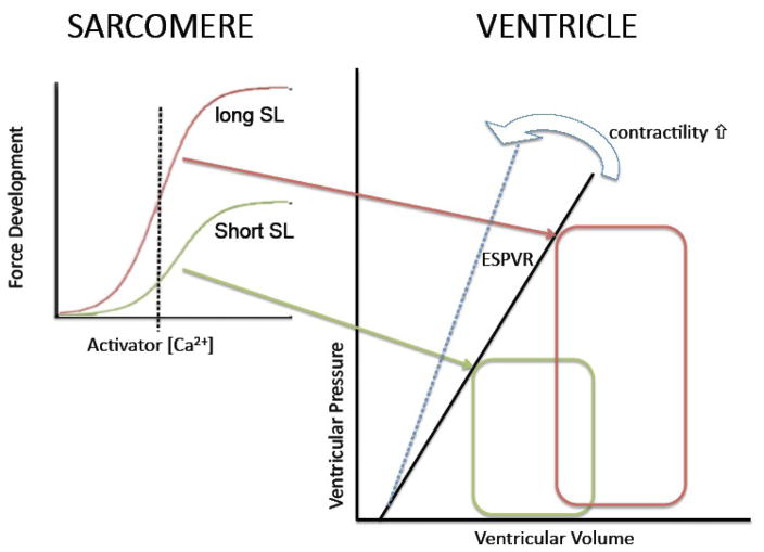
Figure 1. The Frank-Starling mechanism and myofilament length dependent activation.
The Frank-Starling Law of the Heart describes a fundamental property of the heart (figure on the right). That is, for a given contractile state there is a unique relationship between end-systolic pressure reached in the heart and end-systolic pressure (ESPVR); increased contractility results in an increased slope of the ESPVR (cf. blue arrow). Increased ventricular filling (pre-load; red PV loop) leads to an increase in ventricular pressure development at end-systole which allows for i) increased stroke volume for a given systolic pressure (after-load) and ii) sustained stroke volume at elevated systolic pressure. The Frank-Starling mechanism has, as its basis, a modulation of myofilament Ca2+ sensitivity upon a change in sarcomere length as illustrated in the left graphs. Myofilament force development is the result of activation by Ca2+ ions. The relationship between force development and activator [Ca2+] is shifted up and to the left at longer sarcomere length (short SL, green; long SL, red). For a given contractile state (and, thus, cytosolic [Ca2+]; dashed vertical line), more myofilament force is developed at the longer SL (red) leading to a higher ventricular pressure at higher end-systolic volume (red PV loop). Thus, the Frank-Starling Law of the heart is a direct consequence of the myofilament length dependent activation properties of the cardiac sarcomere.

