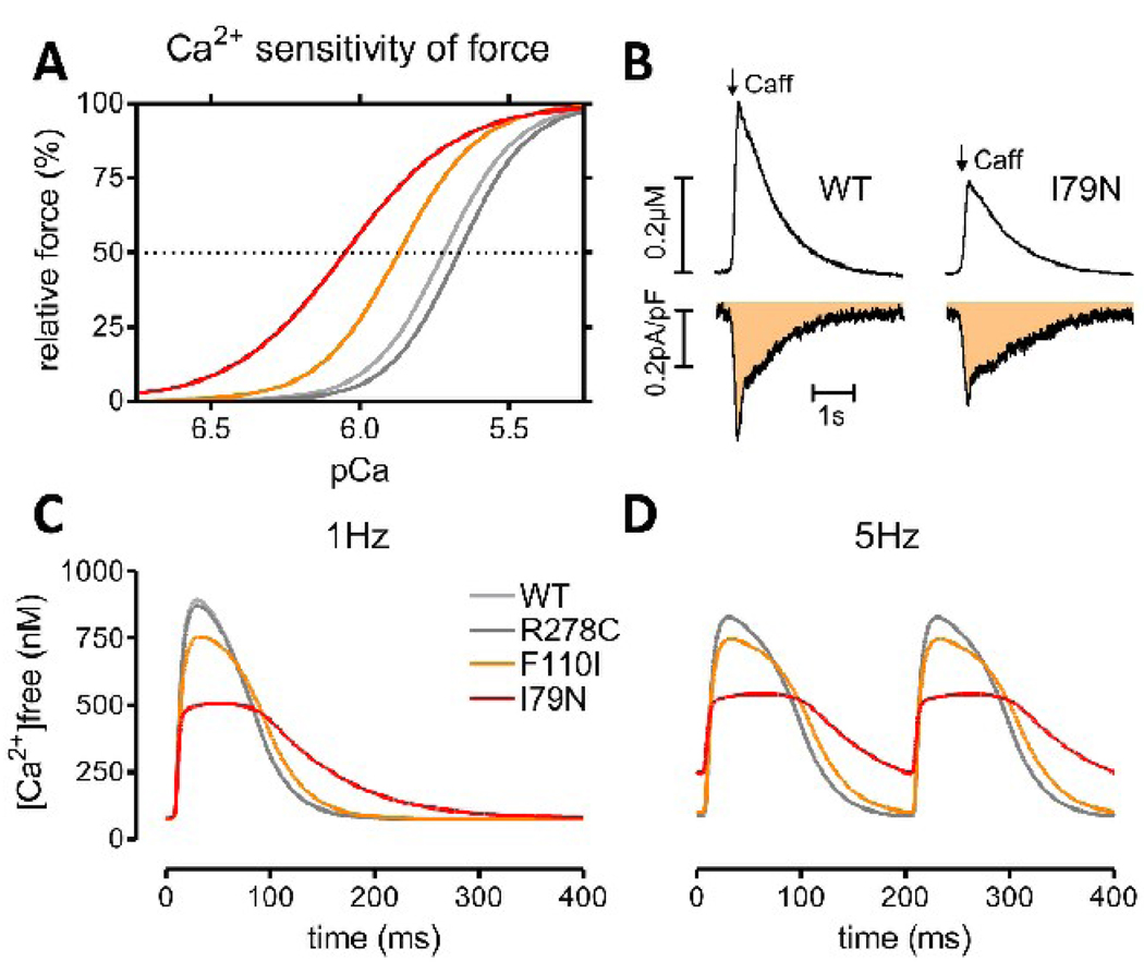
Fig. 1.
The effect of myofilament Ca2+ sensitization on myocyte Ca2+ handling. Panel (A) shows the effect of three TnT mutations on myofilament Ca2+ sensitivity measured in skinned fibers of transgenic mice [78]. Panel (B) shows the effect myofilament Ca2+ sensitization on apparent Ca2+ buffering. The change in total Ca2+ was measured by the Na/Ca exchanger integral (bottom, shaded area) simultaneously with the change in [Ca2+]free (top panel) in voltage clamped fluo-4 loaded myocytes [90, 92]. Note that the peak [Ca2+] is lower in TnT-I79N despite a similar change in total Ca2+, consistent with increased Ca2+ buffering. (C)+(D) Ca2+ transients simulated with LabHEART 5.0 [176] containing a newly integrated force response [177]. Ionic current conductances were adjusted to reproduce a typical mouse action potential. The myofilament Ca2+ sensitivity curves in (A) were reproduced by changing the TnC Ca2+ on/off rates and Ca2+ binding cooperativity. Ca2+ transients are simulated at 1 Hz (C) and 5 Hz (D) pacing frequency. Note that peak [Ca2+] is lower and Ca2+ transients are prolonged in Ca2+ sensitized mutants, while the diastolic [Ca2+] is additionally increased in TnT-I79N at 5 Hz. Panel (C)+(D) were kindly provided by Dr. J. Puglisi, UC Davis.

