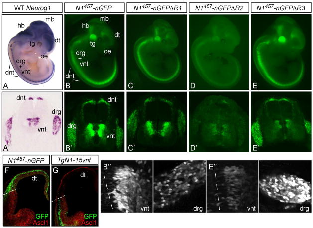
Figure 2. GFP expression from Neurog1-GFP BAC transgenic mice.
mRNA in situ hybridization for Neurog1 in an E11.5 mouse embryo in whole mount (A) or in a cross section of the neural tube (A′). (BE) GFP expression in representative BAC transgenic embryos is shown in whole mount at E11.5. (B′-E′) cross sections show the Neurog1-like expression in the dorsal neural tube (dnt), the ventral neural tube (vnt), and the dorsal root ganglia (drg). (B″,E″) show a higher magnification of the GFP expression in the vnt and drg from N1457-nGFP compared to N1457-nGFPΔR3 to highlight the specific reduction in vnt activity with this deletion. (F,G) telencephalon showing GFP relative to Ascl1 immunofluorescence (red). GFP from N1457-nGFP reflects the endogenous Neurog1 in the dorsal telencephalon while TgN1-15vnt has aberrant expression in the ventral telencephalon. Transgenes are diagramed and results summarized in Fig. 1. dt, dorsal telencephalon; hb, hindbrain; mb, midbrain; oe, olfactory epithelium; tg, trigeminal ganglion.

