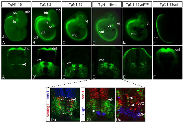
Figure 4. An enhancer for Neurog1-like expression in the ventral neural tube is identified.
(A–F) GFP expression in representative transgenic embryos is shown in whole mount at E11.5. (A′–F′) cross sections show the activity of each transgene in the neural tube. Transgenes are diagramed and results summarized in Fig. 1. Within the spinal neural tube, TgN1-2 directs expression to both dorsal neural tube (dnt) and ventral neural tube (vnt) whereas TgN1-16 and TgN1-13dnt are largely restricted to the dnt (A′, arrowhead indicates the narrow strip of ventral neural tube expression consistently seen with TgN1-16), and TgN1-15 and TgN1-15vnt are restricted to the vnt. Activity for expression in dorsal root ganglia (drg), dorsal telencephalon, olfactory epithelium (oe), trigeminal ganglion (tg), and subsets of hindbrain (hb) and midbrain (mb) is not present in every transgene. For example, drg and dt are largely absent in all transgenes, whereas oe is only seen in TgN1-2, TgN1-15 (tail obscuring oe in this image), and TgN1-15vnt, and tg is only in TgN1-16 and TgN1-2. (Da-Dc) immunofluorescence on E11.5 cross sections of TgN1-15vnt neural tube. (Da) Dashed lines delineate the dorsoventral boundaries of the ventral Neurog1 (red) domain is shared by GFP but also highlight the more dorsal part of this domain is lacking GFP (arrowhead). (Db) Arrows highlight the boundaries of non-overlap of GFP with Ascl1 (red) and Olig2 (blue). (Dc) is a higher magnification of the progenitor domains for V2 interneurons and motoneurons (MN). In the pV2 region there are a few cells that co-express Ascl1 and Neurog1 (Da) and GFP (Dc, arrowheads). Ventral telencephalon (vt) expression, non-Neurog1 pattern, appears in TgN1-15 and its derivatives (see Fig. 2G for a cross section).

