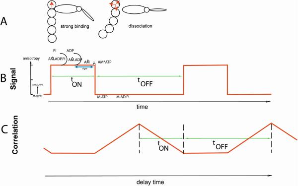
Fig. 9.
The simplest scheme for generating anisotropy fluctuations in contracting myofibrils. A. Actin becomes immobilized when a cross-bridge binds to it resulting in a well defined direction of transition dipole (red arrow, left panel) and high anisotropy. Dipoles become disorganized when a cross-bridge dissociates, leading to low anisotropy (multiple arrows, right panel). B. A simple rectangular signal reflecting binding-dissociation of a cross-bridge. C. Autocorrelation function of a rectangular signal showing duration (tON seconds) of strongly attached state and detached (or weakly attached) state, lasting tOFF seconds.

