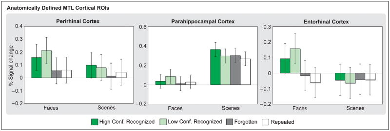
Figure 3.
Encoding activation in anatomically defined MTL cortical regions based on subsequent memory. Percent signal change in perirhinal, parahippocampal, and entorhinal cortices is plotted for stimuli recognized with high confidence (dark green), stimuli recognized with low confidence (light green), forgotten (dark gray), and repeated (white) stimuli for faces and scenes.

