Fig. 5.
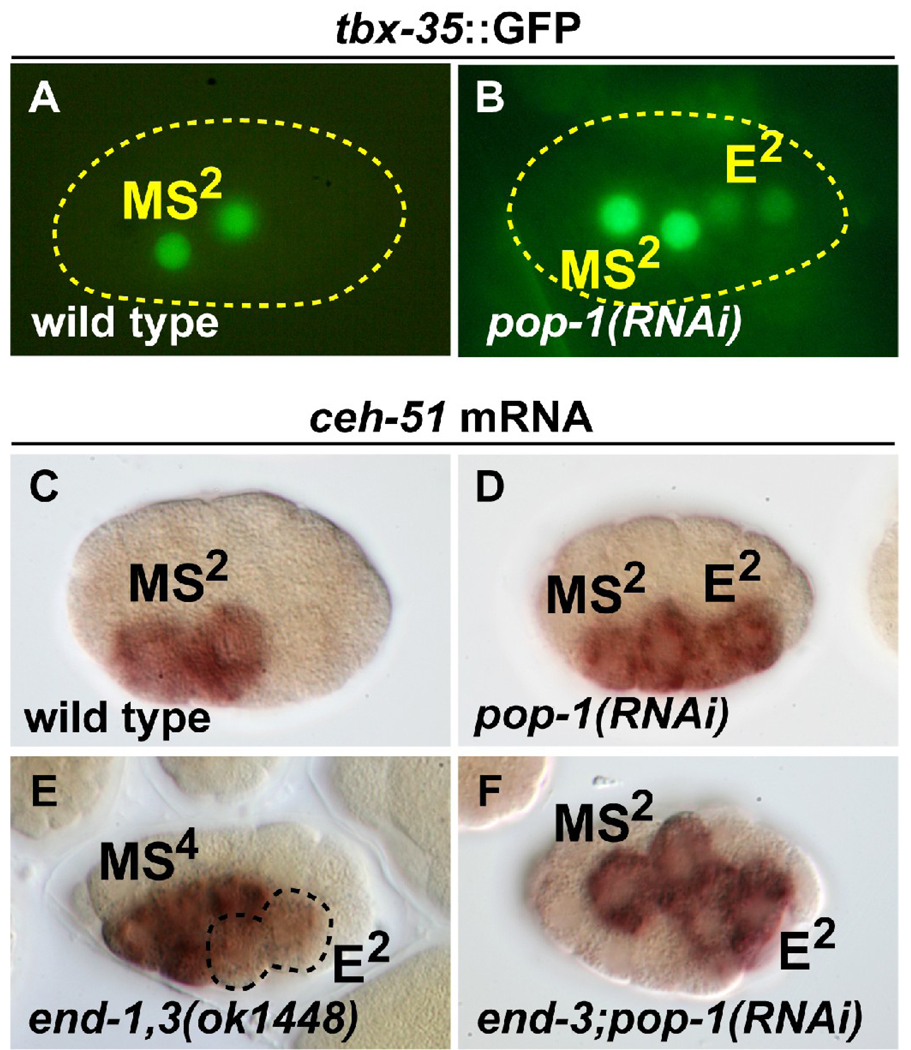
Expression of MS specification factors in pop-1(RNAi) and end-1,3 mutant backgrounds. (A,B) Expression of a tbx-35::GFP reporter in wild-type and pop-1(RNAi) embryos. 100% (n=40) of expressing embryos showed MS lineage-only expression in wild-type, while 12% (n=67) of tbx-35::GFP-expressing pop-1(RNAi) embryos showed ectopic expression in the E lineage as shown; 75% of embryos overall had weak E lineage expression. (C–F) in situ hybridization with a ceh-51 probe. (C) Wild-type staining pattern observed in 91% of embryos in the early MS lineage (MS2 as shown here, or MS4; n=101). (D) Ectopic expression of ceh-51 in the early E lineage was observed in 86% (n=44) of pop-1(RNAi) embryos (the remainder showed either normal expression or did not stain). Results in C and D were previously reported (Broitman-Maduro et al., 2009). (E) Weak ectopic expression of ceh-51 in the E lineage (at E2, as shown here; ectopic expression was also observed at E4), as seen in 41% (n=209) of progeny from end-1(ok558) end-3(ok1448) mothers carrying an end-1,3(+) rescuing array. As this array is transmitted to ~50% of the progeny, ~80% of end-1,3 mutant embryos showed activation of ceh-51 in the E lineage. The remaining embryos showed wild-type expression (MS only). (F) Expression of ceh-51 in MS and E was seen in 88% (n=45) of end-3(ok1448); pop-1(RNAi) embryos. The remaining embryos were either unstained or appeared normal.

