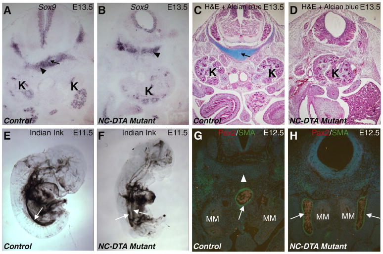
Fig. 4. Disruption of the notochord and floor plate affects chondrogenesis of perinotochordal cells and midline vascular development.
A–B, Sox9 RNA in situ hybridization on transverse sections at the MM level in control (A) and mutant (B) E13.5 embryos. Black arrow in A points to the notochord that is absence in the mutant (B). Black triangles point to the perinotochordal cells positive for Sox9 in both control and mutant samples. C–D, Transverse sections at the MM level in control (C) and mutant (D) E13.5 embryos stained with H&E and Alcian blue (for cartilage). Black arrow in C points to the notochord that is absence in the mutant (D). E–F, India ink injection into the left ventricle of E11.5 control (E) and mutant (F) embryos. The white arrows point to the dorsal aorta. At this time, the control dorsal aorta does not have major branches below the aortic arches (E). However, the mutant aorta is still bifurcated for most of the trunk region (F). G–H, transverse sections at the MM level from control (G) and NC-DTA mutant (H) E12.5 embryos. White arrows point to the αSMA staining (Green) in the aortic wall. White triangle points to the notochord (absent in the mutant). MM is highlighted by Pax2 staining in red. MM: metanephric mesenchyme; K: kidney.

