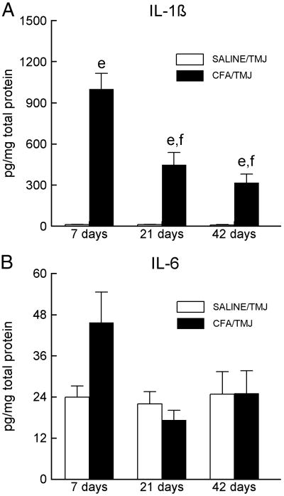
Figure 3.
Interleukin 1β (IL-1β) and IL-6 levels in the TMJ tissues of Sprague-Dawley rats. (A) IL-1β or (B) IL-6 levels within the synovial membrane, joint capsule, retrodiscal tissue, articular disc and a small portion of the lateral pterygoid muscle 7, 21 and 42 days after saline or CFA injection. Within a particular day, the SALINE/TMJ group was compared to the CFA/TMJ group and is indicated by e = P<0.01. The letter “f” indicates that the cytokine levels in the CFA/TMJ-treated groups on days 21 and 42 were significantly (P<0.01) reduced compared to the values of the CFA/TMJ-treated group on day 7. See the legend of Figure 1 for explanations and abbreviations.

