Figure 1.
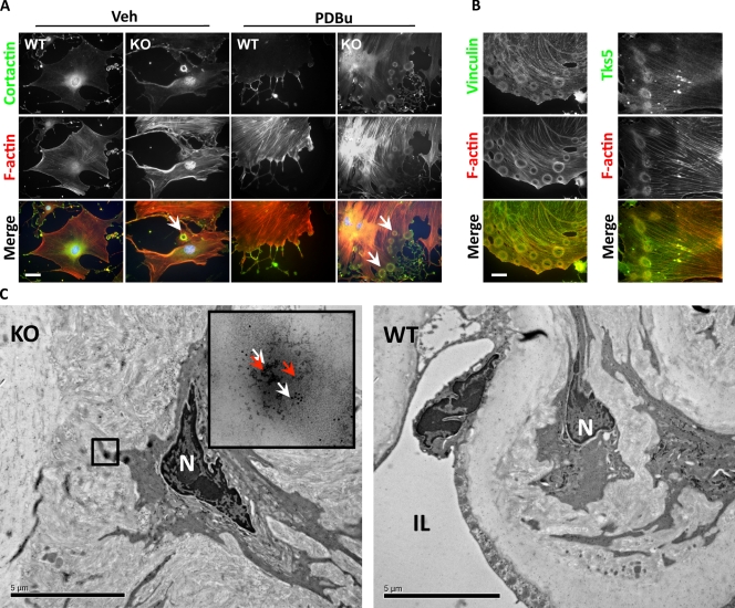
Podosome formation in miR-143(145) KO SMCs in vitro and in vivo. (A) Cortactin and F-actin IF to visualize podosomes in VSMCs isolated from WT and miR-143(145) KO mice treated with PDBu or with vehicle (Veh). Arrowheads indicate the rosettes of podosomes in miR-143(145) KO cells. Quantification of three separate experiments revealed podosomes in 7.6% (±2.5%) of untreated KO SMCs and in 91.3% (±3.2%) of PDBu-treated cells. (B) Colocalization of the podosome proteins vinculin (left) and Tks5 (right) with F-actin in KO VSMCs treated with PDBu. (C) Immunoelectron microscopy of miR-143(145) KO and WT aortas. White arrows indicate cortactin (10-nm gold particles), and red arrows indicate Tks5 (5-nm gold particles). The boxed area indicates what appears to be a rosette of podosomes located in an SMC of the miR-143(145) KO mouse, confirmed by the observation of the gold-labeled podosome markers Tks5 and cortactin in the higher magnification inset. N, nuclei; IL, intima layer. Bars: (A and B) 10 µm; (C) 5 µm.

