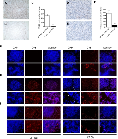
Figure 4.
Long-term cisplatin-treated lung tumors in LSL-KrasG12D/+ mice exhibit enhanced adduct repair in response to a final dose of cisplatin. (A) Representative sensitive lung tumor section (G2) stained with Pt-1,2-d(GpG) from long-term PBS-treated mice given a final 24-h dose of cisplatin. (B) Representative resistant tumor section (G4) from long-term cisplatin-treated mice, treated as in A. (C) Number of Pt-1,2-d(GpG)-positive cells per lung tumor area in long-term PBS-treated (white bar) or cisplatin-treated (black bar) mice given a final dose of cisplatin and sacrificed 24 h later ([***] P < 0.0001). (D) Representative sensitive tumor section (G2) stained with γ-H2AX from long-term PBS-treated mice given a final 24-h dose of cisplatin. (E) Representative resistant tumor (G4) from long-term cisplatin-treated mice, treated as in D. (F) Number of γ-H2AX-positive cells per lung tumor area in long-term PBS-treated (white bar) or cisplatin-treated (black bar) mice given a final dose of cisplatin and sacrificed 24 h later ([***] P < 0.0001). Error bars represent SEM. (G–I). Representative immunofluorescent images of lung tumor sections stained for nuclei (DAPI) or Pt-1,2-d(GpG) (Cy3), and an overlay of these images (Overlay) in mice treated with long-term PBS (LT PBS) or four doses of cisplatin (LT Cis) and given a final dose of cisplatin and analyzed after 0 h (G), 4 h (H), or 24 h (I). The top panels are 10× magnification, and the bottom panels are higher-magnification zooms. (G) Note that adducts persist in normal parts of the lung even after multiple weeks in long-term cisplatin (LT Cis, 0 h). In H (LT Cis, 4 h, DAPI), two tumors are separated by a dotted white line. Approximately 20% of tumors in long-term cisplatin mice had reduced adduct levels as early as 4 h after a final dose of cisplatin (left tumor) whereas the majority of tumors had similar levels of adducts (right tumor) at this time point.

