
Figure 1.
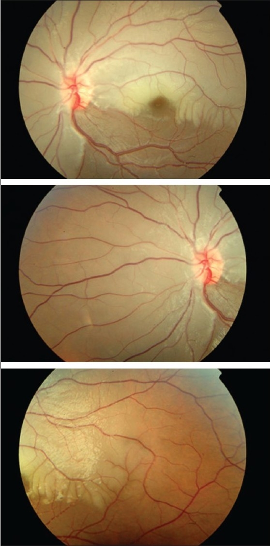
Left eye fundus photograph at presentation showing pallid retinal edema of posterior pole sparing infero-temporal quadrant, arteriolar attenuation and segmentation of blood column in arterioles, suggestive of extensive branch retinal artery occlusion