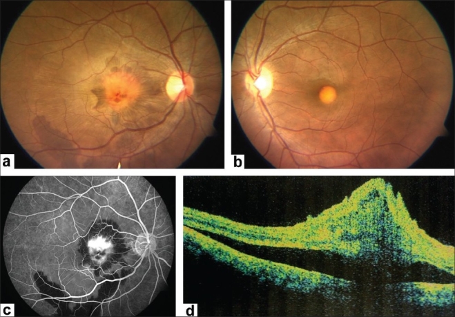
Figure 1.

(At presentation) (a) Fundus examination of the right eye shows a subfoveal vitelliform lesion associated with a yellowish-grey choroidal neovascular membrane, sub-retinal and sub-retinal pigment epithelium hemorrhages extending up to the inferotemporal vascular arcade. Subretinal fluid and internal limiting membrane striae are also noted. (b) Left eye shows an egg yolk appearance at the fovea characteristic of Best's disease. (c) Right eye fundus fluoresceine angiography reveals intense leakage from the choroidal neovascular membrane. Blocked fluorescence corresponds to the subretinal and sub-RPE hemorrhages. (d) Optical coherence tomography of right eye shows presence of SRF, cystoid macular edema, fresh subretinal hemorrhage and disorganized RPE-choriocapillaris complex suggestive of choroidal neovascular membrane
