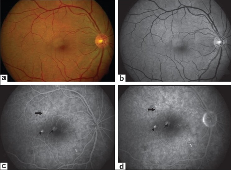
Figure 1.

(a and b) Fundus and red-free photograph of the right eye showing mottled appearance superotemporal to the fovea. (c and d) Angiogram during transit and late phase showing hyperfluorescence suggestive of pigment epithelium detachment (arrows). Additionally, an area of punctate hyperfluorescence (solid arrow) is shown with an absence of smoke stack or subretinal fluid
