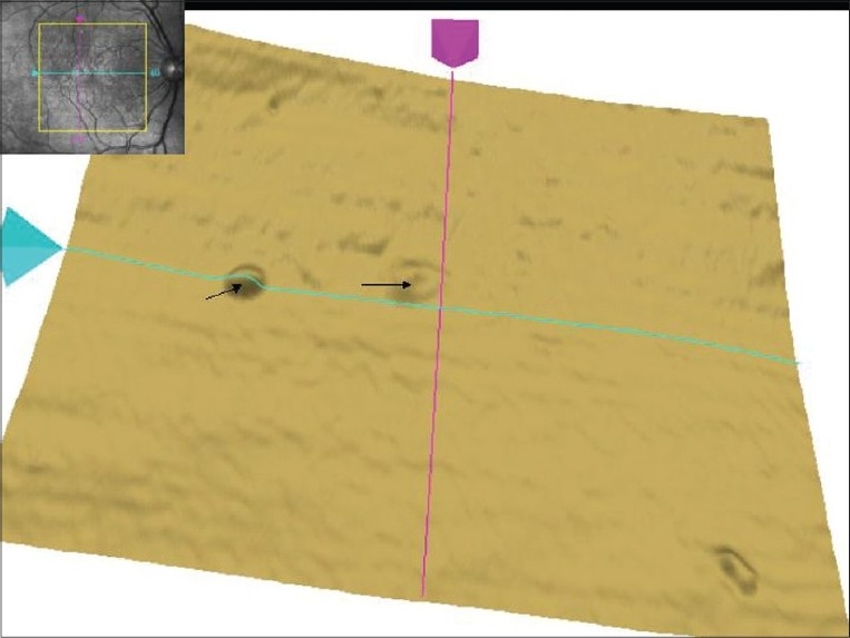
Figure 3.

Single layer retinal pigment epithelium scan of right eye on Macular Cube 512 × 128 Combo shows pigment epithelium detachment s (arrows). In addition, the superior part of retinal pigment epithelium layer shows uneven surface with bumps. The inset shows fundus image with scan cube overlay
