
Figure 5.
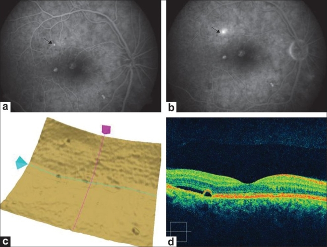
(a and b) Same eye(right eye) 3 months later shows expanding dot sign on fluorescein angiography (arrows). (c). Single layer RPE scan shows more uneven and bumpy surface compared to previous scan in Figure 2. (d). Raster Line scan right eye shows pigment epithelium detachment with subretinal fluid