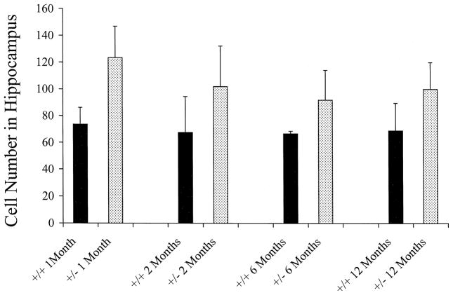
Fig. 5.
Increased numbers of GFAP-positive astrocytes in hippocampus in Nf1/nf1 mice at different ages. GFAP-positive astrocytes were counted in sections through the hippocampus of 1 month, 2 month, 6 month and 1 year old animals. Counts from individual animals (2 sections/animal) were pooled for each age group (n =6 animals each age and each genotype). Error bars represent standard deviation. Statistical significance was observed for all ages (see text).

