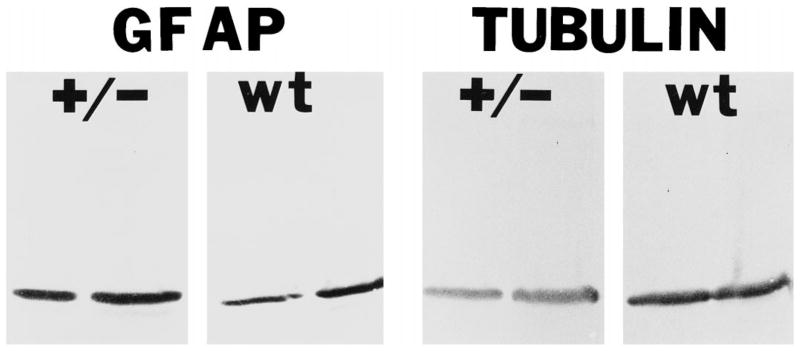
Fig. 7.
GFAP protein is increased in Nf1/nf1 brain lysates. GFAP and tubulin levels were quantified with Western blot analysis. 3 μg (left lane in each set) and 10 μg (right lane in each set) of cytoskeletal extracts from Nf1/nf1 brains (+/−) or wild type (wt) brains were transferred to nitrocellulose and probed with antibodies recognizing GFAP (left four lanes) or tubulin (right four lanes). Nf1/nf1 brains have approximately two-fold increased levels of GFAP proteins.

