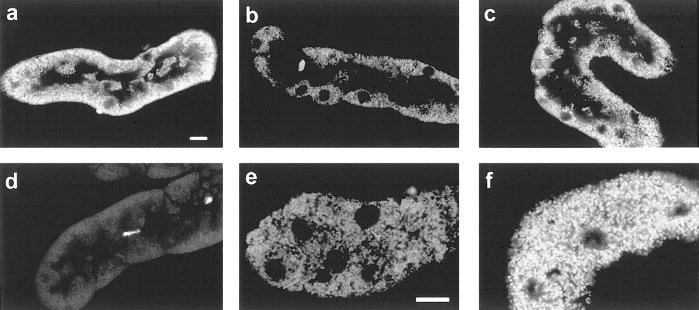
Fig. 4.
Mitochondrial tetramethylrhodamine methyl ester (TMRM) uptake. Tubules were stained with TMRM and propidium iodide after: oxygenated control incubation (a); 60 min hypoxia followed by 60 min reoxygenation with no extra substrates (b, e) or with 4 mM α-KG/ASP during reoxygenation (c, f); or 5 μM FCCP + 5 mM glycine for 15 min (d). Optical sections were viewed at 568 nm excitation, 585 nm emission. Panels a–d are cuts through the midplanes of the tubules showing the basally oriented mitochondria around the periphery of the tubule profile. The unstained areas in the center of each tubule are the apical portions of the cells, which are devoid of mitochondria, and the tubule lumens. Nuclei are seen as the circular areas devoid of staining. The full tubule profile in panel d occupies approximately the same area and is similarly oriented to those in the other panels, but is poorly seen because the mitochondria are unstained. The brightly stained circular structures are the rare propidium iodide-positive nuclei that were no more frequent than those seen in controls. Panels e and f are higher magnification cuts through the basal sections of the cells to show mitochondria at higher resolution. Bars in panels a and e are 10 μm. Results are representative of 8–10 tubules from 4–5 different preparations viewed under each experimental condition.

