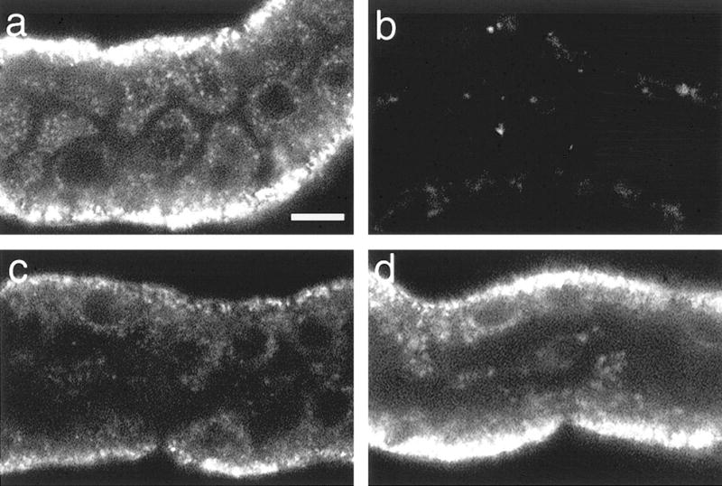
Fig. 6.

Confocal microscopy of red JC-1 aggregates after hypoxia plus reoxygenation. Tubules were stained with JC-1 after: oxygenated control incubation (a); 5 μM FCCP + 5 mM glycine for 15 min (b); or 60 min hypoxia followed by 60 min reoxygenation with no extra substrates (c) or with 4 mM glutamate + malate during reoxygenation (d), then viewed by confocal microscopy at 568-nm excitation, 585-nm emission. Magnifications are the same for all panels. Bar: 10 μm. Unavoidable photobleaching results in dropout of the signal from the most severely affected mitochondria in the no extra substrate group.
