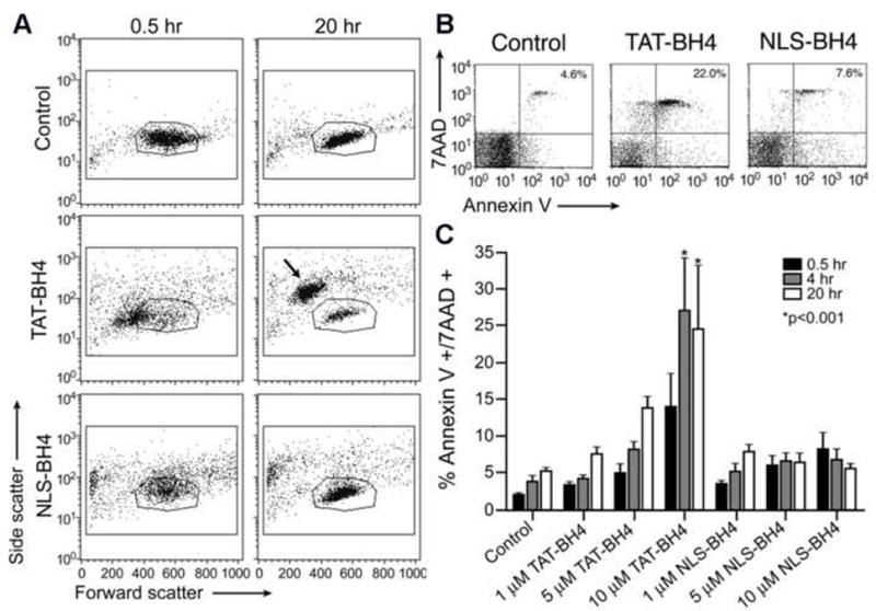
Figure 2.

Flow cytometric assessment of PBMCs treated with CPP-BH4 conjugates. (A) Morphological changes in human PBMCs following treatment with PTD-conjugated BH4 peptides. These data show the entire population of isolated PBMCs from one representative donor (no gating was done). The outlined region (polygon) indicates the typical FSC/SSC region where unmanipulated lymphocytes are observed. (B and C) Morphological changes correlate with phosphatidylserine exteriorization and loss of membrane integrity following peptide treatment. (B) Representative FACS plots 4 h after the administration of saline, NLS-BH4, or TAT-BH4 (10 μM). These data show the entire population of isolated PBMCs from one representative donor (no gating was done). (C) The time and dose dependence of Annexin V and 7AAD staining of PBMCs following peptide exposure. These data are calculated from the percentage of cells in the upper right quadrant (AnxV+/7AAD+) of FACS diagrams as shown in panel A (N=5). Statistical significance marked is compared to untreated (control) cells at the same time point.
