Figure 3.

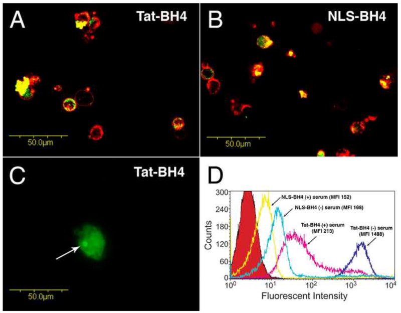
Uptake of fluorescein-conjugated Tat-BH4 and NLS-BH4 in human peripheral mononuclear cells. Panels A and B show, by confocal microscopy, representative images (400X) of the intratracellular localization of both fluorescein-labeled Tat-BH4 and fluorescein-labeled NLS-BH4 as indicated. Cells were incubated with the fluorescein-labeled peptides in the absence of serum (5μM, 1h, green color) and then treated with Alexa 594-conjugated Wheat Germ Agglutinin (red color) to distinguish the plasma membrane. Gain and photomultiplier settings were held constant across samples. Panel C shows, by fluorescence microscopy, a representative image (600X) of the nucleolar accumulation (arrow) of internalized fluorescein-labeled Tat-BH4 (5μM, 1h, serum-free). Panel D shows, by flow cytometry, the fluorescent intensities of cells treated with fluorescein-conjugated Tat-BH4 and NLS-BH4 (5μM) in both the presence and absence of serum as well as untreated cells (red peak).
