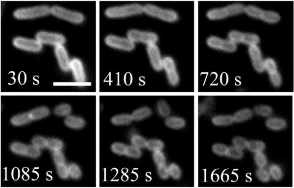
Fig. 13.
Binary fission of E. coli cells stained with probe 8. The cells were imaged every 5 s for 30 min by using fluorescence microscopy. The scale bar represents 2 μm. Reproduced with permission from ref. 21. Copyright Wiley-VCH Verlag GmbH & Co. KGaA.

