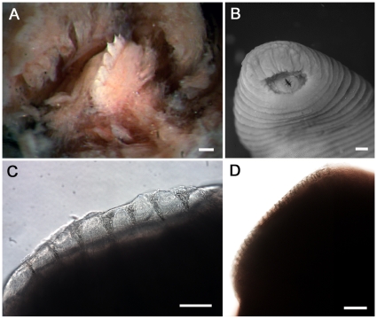
Figure 2. Comparative jaw morphology of Tyrannobdella rex.
(A) Stereomicrograph of the single dorsal jaw of T. rex with large teeth. Scale bar is 100 µm. (B) Tyrannobdella rex anterior sucker exhibiting velar mouth and longitudinal slit through which the dorsal jaw protrudes when feeding. Scale bar is 1 mm. (C) Compound micrograph in lateral view of eight large teeth of T. rex. Scale bar is 100 µm. (D) Lateral view of jaw of Limnatis paluda illustrating typical size of hirudinoid teeth. Scale bar is 100 µm.

