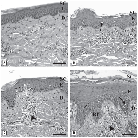
Figure 3.
Light micrographs showing effects of 20-nm washed Ag-nps on porcine skin (stained with hematoxylin and eosin). (A) Untreated control. Skin exposed to 0.34 μg/mL (B); 3.4 μg/mL (C); and 34 μg/mL (D) 20-nm washed Ag-nps. Abbreviations: D, dermis; E, epidermis; RP, rete peg; SC, stratum corneum. Large arrows indicate intracellular epidermal edema; small arrows, focal areas of intercellular epidermal edema; arrowheads, perivascular inflammation. Bars = 60 μm.

