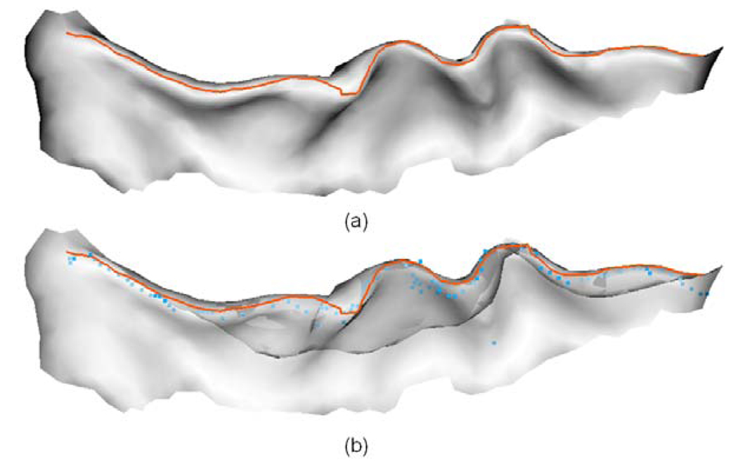
Fig. 17.
An example of comparison of sulcal fundi extraction results between our results with BrainVISA’s results. The orange curves indicate the extracted central sulcal fundi by our method, and the blue points represent the sulcal fundi points generated by the BrainVISA. The sulcus is viewed from inside to outside. (a) Our extracted sulcal fundi overlaid on the central sulcus. (b) Our extracted sulcal fundi and BrainVISA generated sulcal fundi points overlaid on the central sulcus, where the opacity of the surface is set to be 0.6 for the convenience of inspection.

