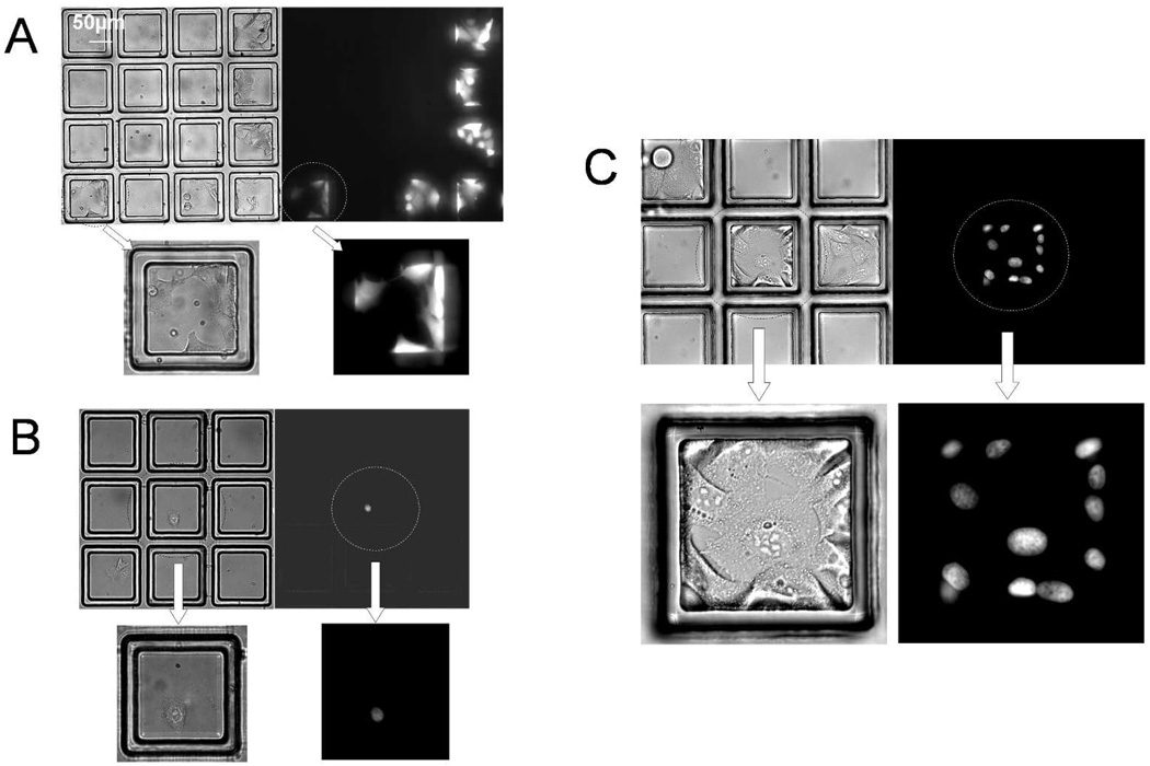
Figure 4.
Testing cell viability and growth on microcup arrays. (A) Test of cell viability: brightfield and fluorescence images of wild type Hela cells cultured on a microcup array for 72 hr and then labeled with Calcein Red-Orange. (B–C) Test of cell replication: brightfield and fluorescence images of HeLa cells captured in microcups 1 hr after plating (B) and after 96 hr in culture (C). (B) The cell in the center microcup possesses a fluorescent nucleus due to stable expression of a GFP/histone-H1 fusion protein. (C) Images of a region of the microcup array containing a clonal colony of GFP/histone-H1-expressing HeLa cells and colonies of non-fluorescent cells in adjacent microcups.

