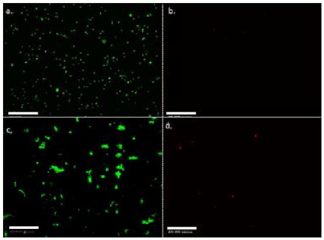
Figure 5.

Fluorescent images of living (a) and dead (b) cells exposed to no sound field or UV-light in cell growth media; images of living (c) and dead (d) cells exposed to 20 min of 2.32 MHz ultrasound at and 4 VRMS with no UV-light. Images indicated that there was no significant cell death from application of the sound field. The cell concentration was equal to 5 × 10⁶ per ml of fluid. The scale bar is equal to 500 μm.
