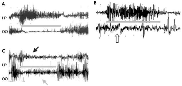
Figure 1.
EMG of the levator palpebrae (LP) and orbicularis oculi (OO) in the normal individual (A), in the MSA-p patient with blepharospasm (B) and in the patient with blepharospasm and apraxia of eyelid opening (C). Comparing with normal finding (A), EMG showed a marked contraction of OO (white arrow) during eye opening (gray line) (B). During eye opening, OO inhibition was incomplete (gray arrow) and LP contraction was not sustained (black arrow) (C).

