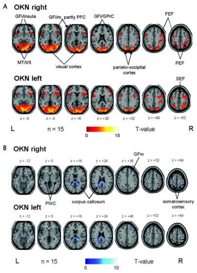
Figure 5.
(A) OKN during rightward (upper row) and leftward (lower row) small-field visual stimulation vs. rest condition (stationary screen) in a group of 15 healthy right-handed volunteers elicited very similar bilateral activations of the visual cortex, which merged into the adjacent occipitotemporal (motion-sensitive area MT/V5) and parietooccipital areas including the parietal eye field (PEF) along the intraparietal sulcus. Additional activations were located nearly symmetrically in the anterior insular region and the adjacent parts of the inferior frontal gyri (GFi) as well as in different ocular motor structures such as the prefrontal cortex (PFC, GFm=middle frontal gyrus), frontal (FEF), and supplementary eye fields (SEF). For illustrative purposes, voxels above a threshold of P≤0.005 uncorrected are shown. (B) OKN during rightward (upper row) and leftward (lower row) small-field visual stimulation in a group of 15 healthy right-handed volunteers caused deactivations in the posterior corpus callosum which partly merged into adjacent parts of the posterior cingulate gyrus and optic radiation. Additional bilateral deactivations were found in the parieto-insular vestibular cortex (PIVC) in the posterior insula, in the central sulcus region (best attributed to the somatosensory cortex), and in the frontal-most and medial part of the right middle frontal gyrus (BA 8, GFm). For illustrative purposes, voxels above a threshold of P≤0.005 uncorrected are shown.92

