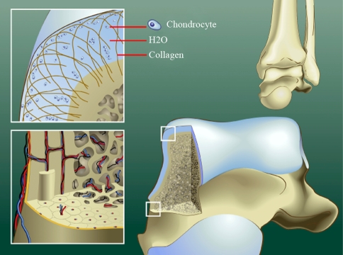
Fig. 1.
Schematic diagrams showing normal anatomy of ankle cartilage, subchondral plate and subchondral bone area. The cartilage consist of chondrocytes that lie groupwise in lacunae of the extracellular matrix, which contains collagen fibers in an arcwise configuration, hyaluronic acid, proteoglycans and 75% water (upper left). The hollow haversian canal that runs longitudinally down the center of the osteon in compact bone contains an arteriole, venule and lymphatic duct for vascular and lymphatic drainage. The Volkmann canals run perpendicular to and connect the Haversian canals (lower left)

