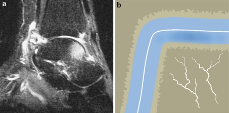
Fig. 8.
a Sagittal T2-weighted MRI study of an ankle with a reticular bone bruise. The white area in the anterior talus represents bone edema. b Schematic diagram of a reticular bone bruise with intact subchondral bone plate. This type of bone bruise heals from the periphery to the center without complications

