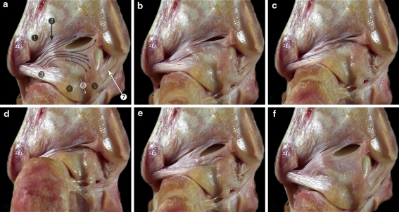
Fig. 11.
Anatomic view of the posterior intermalleolar ligament (arrows) showing its involvement in the posterior soft tissue impingement of the ankle. From dorsiflexion (a) to plantar flexion (d), to dorsiflexion (f). 1 Superficial component of the posterior tibiofibular ligament; 2 deep component of the posterior tibiofibular ligament or transverse ligament; 3 posterior talofibular ligament; 4 lateral talar process; 5 medial talar process; 6 tunnel for the flexor hallucis longus tendon; 7 deep layer of the medial collateral ligament (deep posterior tibiotalar ligament)

