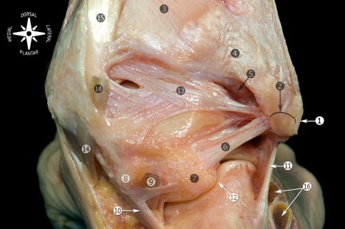
Fig. 8.
Posterior view of the anatomic dissection of the ankle ligaments. 1 Tip of the fibula; 2 peroneal groove of the fibula; 3 tibia; 4 superficial component of the posterior tibiofibular ligament; 5 deep component of the posterior tibiofibular ligament or transverse ligament; 6 posterior calcaneofibular ligament; 7 lateral talar process; 8 medial talar process; 9 tunnel for flexor hallucis longus tendon; 10 flexor hallucis longus retinaculum; 11 calcaneofibular ligament; 12 subtalar joint; 13 posterior intermalleolar ligament; 14 flexor digitorum longus tendon (cut); 15 tibialis posterior tendon; 16 peroneal tendons

