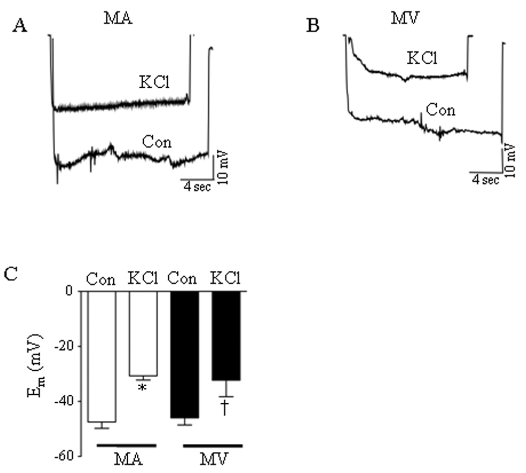
Figure 1.
Depolarization of rat mesenteric arteries (MA) and veins (MV) by 60 mmol/L KCl. (A, B) Membrane potential tracings in isolated, pressurized mesenteric arteries (MA) and mesenteric veins (MV), respectively, under control (Con) drug-free conditions and after incubation with KCl. (C) Resting membrane potential and amplitude of KCl-induced depolarization was not significantly different between MA and MV (n = 4–6). * = significant difference between Con and KCl (MA), † = significant difference between Con and KCl (MV) (p<0.05).

