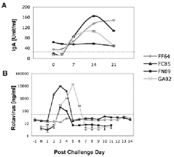
Figure 1.

Rotavirus plasma IgA (A) and fecal shedding (B) are shown upon experimental challenge of 4 juvenile macaques with human rotavirus. Dotted lines represent cut-offs between negative and positive measurements—as previously established by average values corresponding to populations of control (negative and positive) macaques. Immunoassays used to measure the rotavirus plasma IgA and fecal shedding were described in detail elsewhere (Sestak et al. 2004; McNeal et al. 2005; Zhao et al. 2005).
