Figure 2.
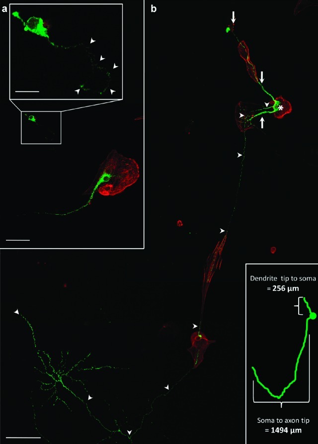
Cell-to-cell contact improves magnocellular neuron development. Neurons cultured on laminin show improved development when in contact with support cells rather than laminin alone. (a) Magnocellular neuron (green) on laminin alone and adjacent to magnocellular neuron on support cell (red). Magnified inset shows stunted magnocellular neuron growth; arrowheads identify the axon. (b) Extensive development of magnocellular neurons in low-density, serum-free culture for 8 d. The neurophysin-labeled magnocellular neurons (green) with soma (∗), axon (arrow heads), and dendrites (arrows), are situated on support cells (red), which are easily identified with rhodamine phalloidin. Distal dendrite tip to distal axon tip measurement = 1750 μm. Distance for distal dendrite to soma (∗) is 256 μm and for soma to axon tip is 1494 μm. Scale bars: top = 15 μm; center = 50 μm; bottom = 100 μm.

